15 Body Parts Named After People
Curious thing about our bodies – they hide odd histories inside. Take how often bits are tagged with a person’s surname.
Back then, researchers loved attaching names to what they found, either theirs or a fellow scholar’s. Those labels just never left, outliving the very people who gave them.
Even now, old titles linger where bones meet nerves. Now here’s a thought – those labels doctors toss around aren’t just random words.
A few ring familiar during regular checkups. Others stay hidden until trouble appears on test results.
Behind each one sits someone who spent years figuring things out, often without applause.
Achilles Tendon

Achilles tendon links calf muscle to heel bone – it ranks among the toughest tendons people have. Named after a legendary Greek fighter said to be vulnerable only at his heel.
Back in the 1600s, Philip Verheyen gave it that title following a close look at his own severed limb, then wrote down what he saw. Hidden within each person’s foot lies this tiny echo of an ancient story.
Fallopian Tubes

Eggs move from ovaries to womb through the Fallopian tubes, while their name honors Gabriele Falloppio, an Italian scholar of the 1500s. Though he wasn’t the first to spot these structures, his written accounts stood out for how deeply they went into detail.
Based in Padua and later working in Pisa, he mapped parts of human reproduction with unusual care. Because of those records, people began linking his name to the tubes without question.
Medical training even now carries ideas rooted in what he laid down centuries ago.
Eustachian Tube
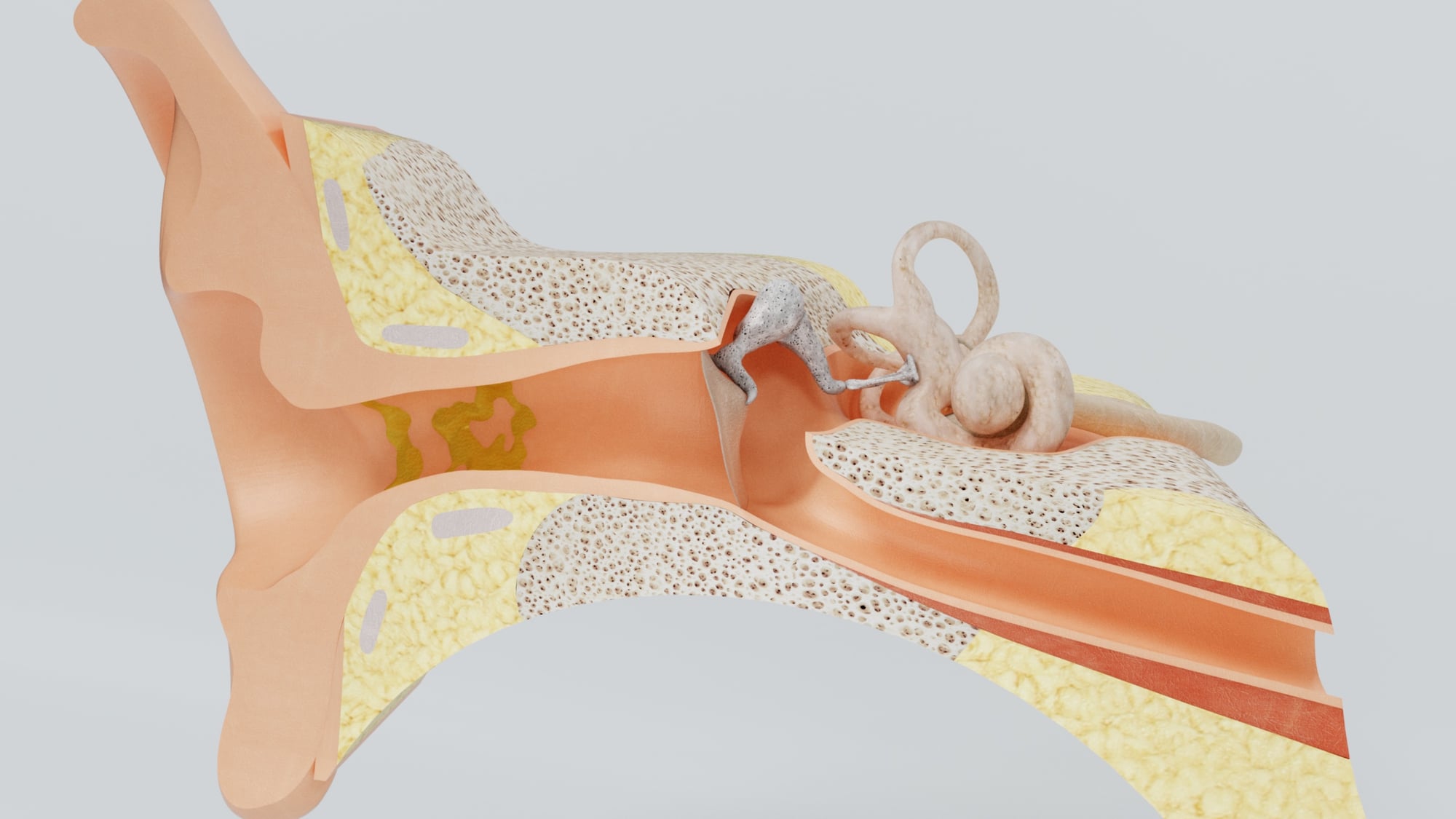
From the middle of your ear down toward the throat’s rear stretch a tiny passage known mostly when airplane travel makes hearing shift suddenly. This feature bears the label of Bartolomeo Eustachi, who studied body structure centuries ago in Italy.
Though he worked long before modern tools existed, his sketches captured details few others matched at the time – some unseen by the public until years passed him by. Glands near kidneys caught his eye too; so did parts inside those organs themselves.
Yet one small channel tucked near the head remains what links his life to today.
Adam’s Apple
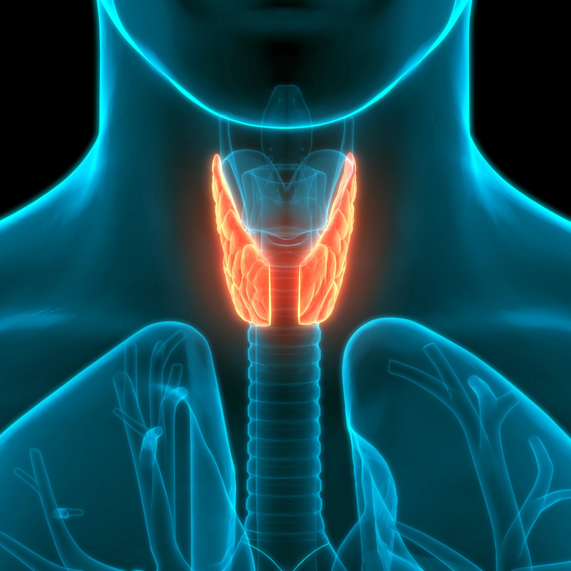
That lump on the neck? It belongs to the thyroid cartilage shaping the voice box. Called the Adam’s apple due to an old tale – some say fruit lodged in Adam’s throat long ago.
Truth be told, scripture shaped the label far more than biology ever did. Over time though, doctors and everyday speech kept the phrase alive.
When boys go through changes, male hormones steer cartilage into sharper angles. Because of this shift, their outline often shows up clearer.
Bartholin’s Glands

One side of the vaginal entrance holds a Bartholin gland, another sits on the opposite side – each makes moisture for smoother movement. A doctor from Denmark, Caspar Bartholin the younger, put these into writing back in sixteen seventy-seven.
His father also studied bodies, so science ran deep in their household through years of work. Tiny things, easily forgotten, slip minds until something goes wrong like clogging or swelling.
Cowper’s Glands
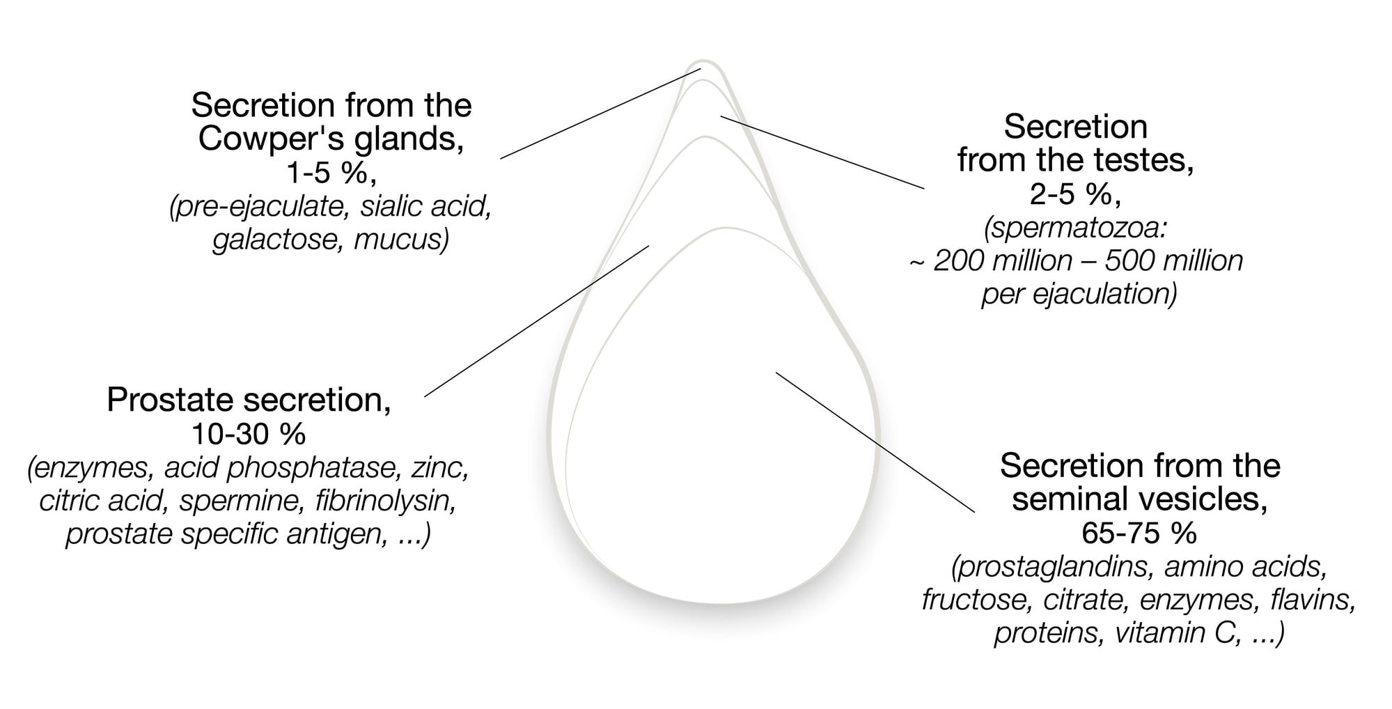
Down near the male prostate sit two tiny organs known as Cowper’s glands. These release a prep liquid ahead of other fluids moving through the urethra.
A man called William Cowper, a doctor in England, wrote about them back in 1699. Before him though, someone else – Mery, a Dutch anatomy expert – had already pointed them out.
That caused some argument over what to call the structures. In time, Cowper’s label stuck, despite others having spotted them too.
Islets Of Langerhans
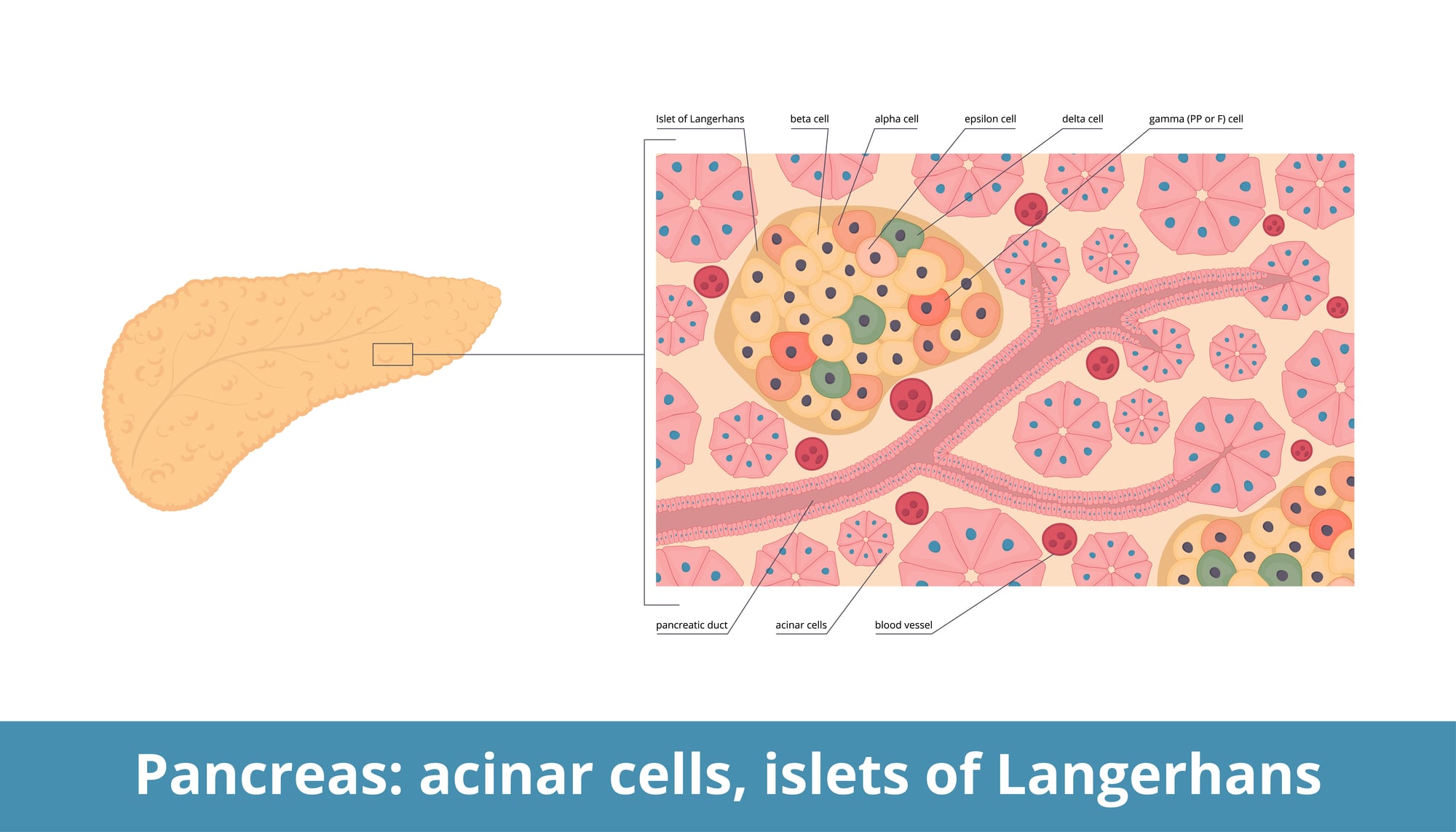
Tiny cell groups in the pancreas make insulin and glucagon, helping control blood sugar levels. Found by Paul Langerhans – then only twenty-two – in 1869 during his studies in Germany.
Back then, their purpose stayed unclear until later research tied them to diabetes. While still a young student, he uncovered something lasting in medical science.
A rare achievement at such an age leaves behind a quiet legacy.
Broca’s Area

Broca’s area is a region in the left hemisphere of the brain that handles speech production. It was identified by French surgeon Paul Broca in 1861 after he studied a patient who could understand language but could not speak.
The patient, known in medical history as ‘Tan’ because that was the only word he could say, helped Broca pinpoint exactly where in the brain language originates. It was one of the first times scientists proved that specific functions live in specific parts of the brain.
Wernicke’s Area

While Broca’s area handles speech production, Wernicke’s area handles language understanding, and the two work closely together. Carl Wernicke, a German physician, identified this region in 1874, just a few years after Broca’s discovery.
Patients with damage to Wernicke’s area can speak fluently but produce sentences that make no sense, a condition now called Wernicke’s aphasia. Wernicke made this discovery at just 26 years old, which makes it even more impressive.
Poupart’s Ligament
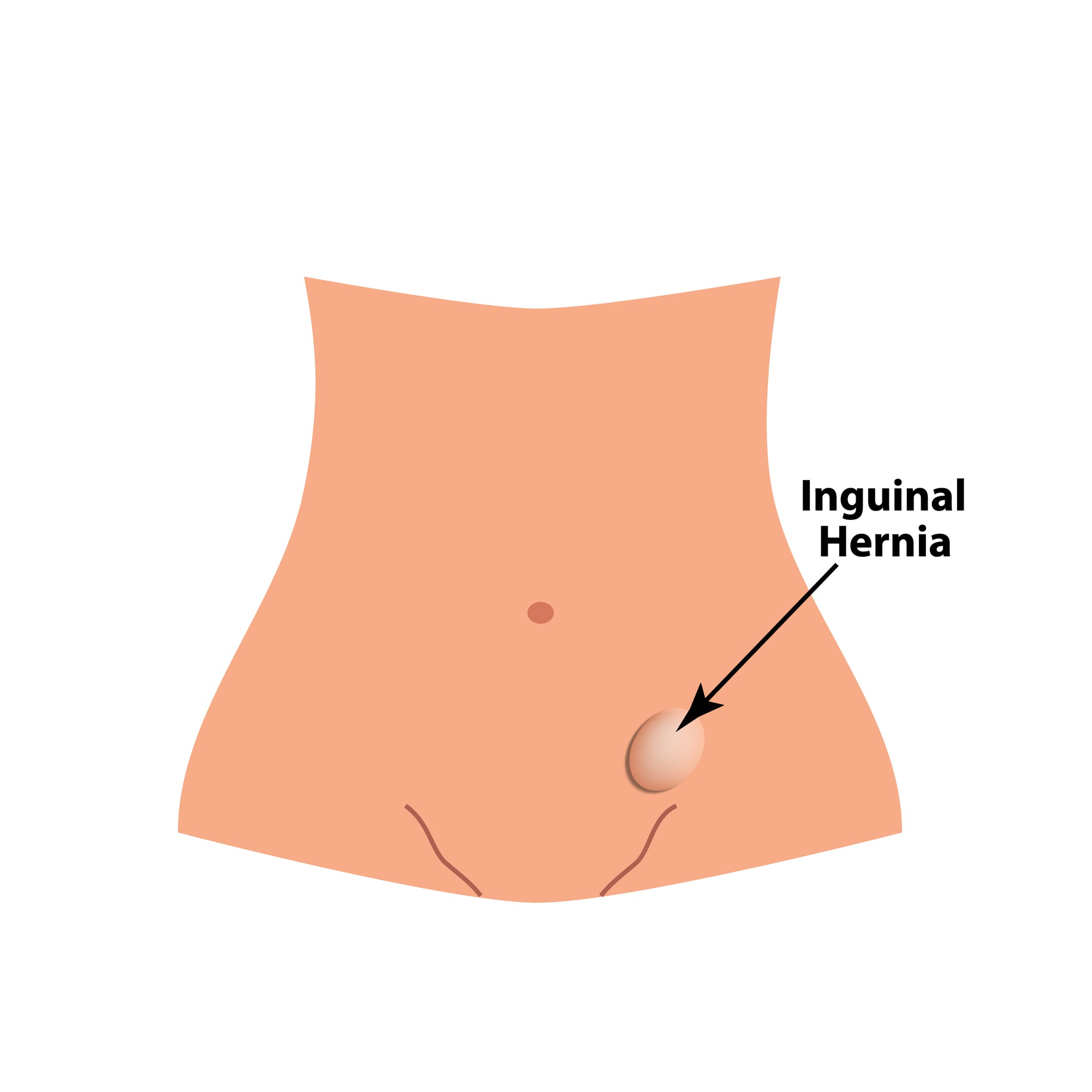
Poupart’s ligament, more commonly called the inguinal ligament, runs from the hip bone to the pubic bone and forms the base of the abdominal region. François Poupart, a French surgeon and naturalist, described it in detail in the late 17th century.
The ligament is important in surgical procedures involving hernias, particularly inguinal hernias that occur in the groin area. Poupart also had a strong interest in natural history and insects, which made him a rather unusual combination of anatomist and scientist.
Bundle Of His
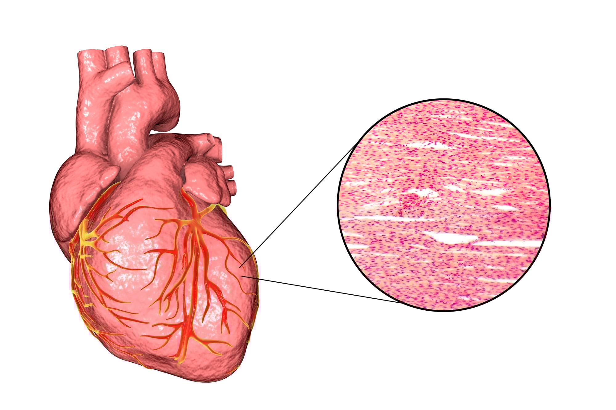
The Bundle of His is a group of heart muscle cells that carry electrical signals from the upper chambers of the heart to the lower chambers, helping to coordinate the heartbeat. Wilhelm His Jr., a Swiss cardiologist, discovered it in 1893.
Without this bundle functioning correctly, the heart can develop a condition called heart block, where the upper and lower chambers beat out of sync. His father, Wilhelm His Sr., was also a respected anatomist, making the His family something of a medical dynasty.
Circle Of Willis

The Circle of Willis is a ring of connected arteries at the base of the brain that supplies blood to much of the brain and surrounding structures. Thomas Willis, an English physician, described it in 1664 in his landmark book on the anatomy of the brain.
He worked alongside the architect Christopher Wren, who drew the detailed illustrations for the book, making it one of the most beautifully documented medical texts of its time. The circle acts as a backup system, rerouting blood if one artery becomes blocked.
Stensen’s Duct

Stensen’s duct is the channel through which saliva travels from the parotid gland into the mouth. Niels Stensen, a Danish scientist, discovered it in 1660 at the age of just 22.
Stensen went on to make contributions in geology and theology as well, and he eventually became a Catholic bishop. Despite his many achievements, it is this small saliva duct that most people associate with his name in medical textbooks.
Pouch Of Douglas
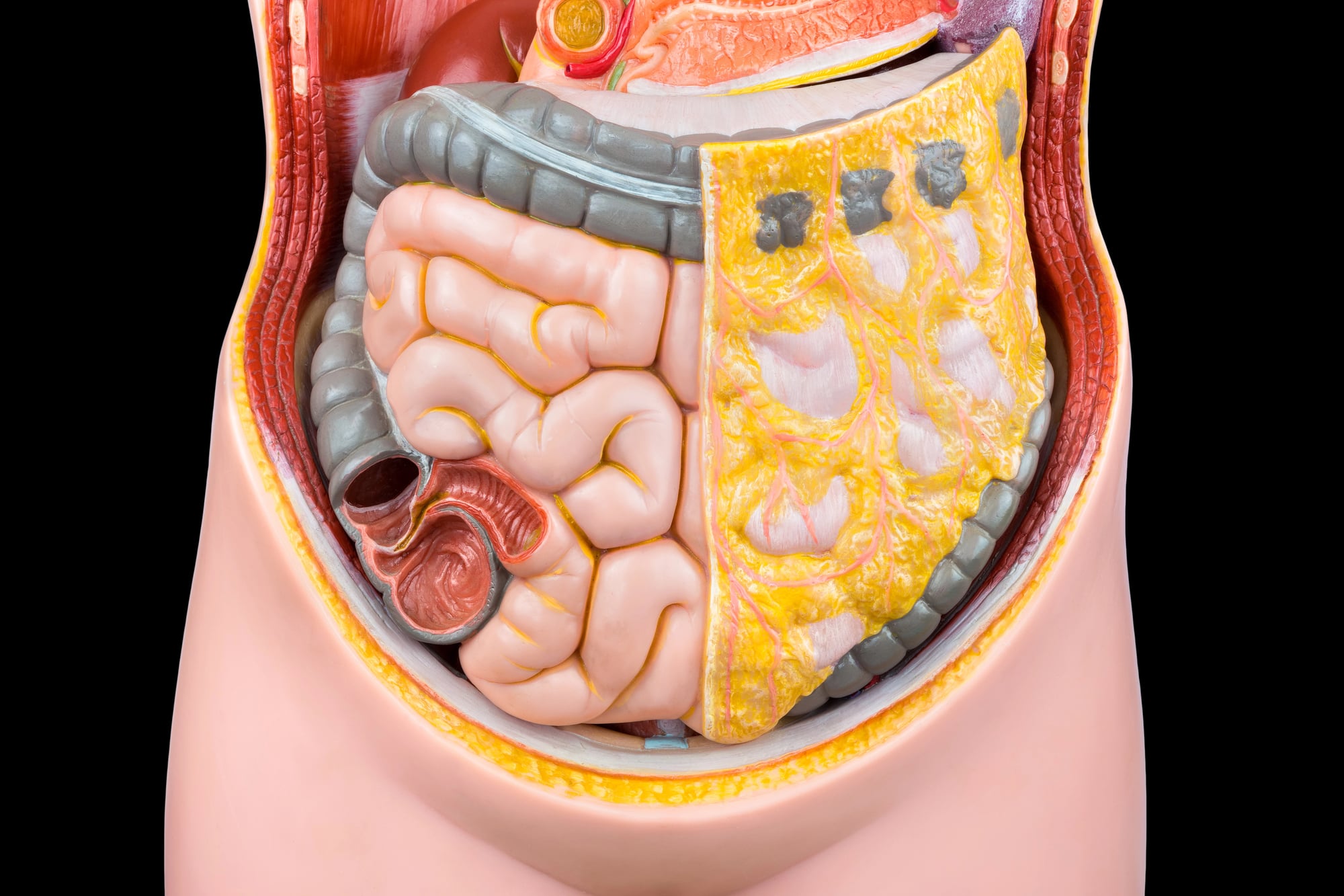
The Pouch of Douglas is a space in the lower abdomen between the uterus and the rectum in women. It was named after James Douglas, a Scottish anatomist who described it in the early 18th century.
This pouch is clinically significant because fluid, blood, or abnormal cells can collect there, making it an important area to examine during certain medical procedures. Douglas also described several other anatomical structures, but this particular pouch became his most lasting contribution.
Haversian Canals

Haversian canals are tiny channels inside bone tissue that carry blood vessels and nerves through the hard outer layer of bone. Clopton Havers, an English physician, described them in his 1691 work on bone structure.
Before his research, the internal structure of bone was not well understood, and his observations helped establish the field of bone histology. These canals are so small they can only be seen under a microscope, yet they keep bone tissue alive by supplying it with nutrients constantly.
Their Names, Still Working

It is worth stopping to appreciate how a doctor’s observation made in a cold 17th-century laboratory still shows up in a modern hospital report today. Many of these people worked without advanced tools, relying on careful eyes, basic instruments, and a lot of patience.
Their names attached to the body are not just labels; they are quiet reminders that science moves forward one curious person at a time. The body carries history, and some of that history has a name.
More from Go2Tutors!

- The Romanov Crown Jewels and Their Tragic Fate
- 13 Historical Mysteries That Science Still Can’t Solve
- Famous Hoaxes That Fooled the World for Years
- 15 Child Stars with Tragic Adult Lives
- 16 Famous Jewelry Pieces in History
Like Go2Tutors’s content? Follow us on MSN.